NIH grant to help engineers visualize Alzheimer’s
From the WashU Newsroom…
With support from the National Institutes of Health (NIH), an engineer at Washington University in St. Louis plans to push the envelope of microscopic imaging to better visualize the molecules involved in Alzheimer’s disease.
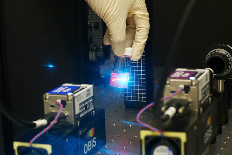
Matthew Lew, assistant professor of electrical & systems engineering in the School of Engineering & Applied Science, recently won a five-year, $1.6 million Maximizing Investigators’ Research Award from the NIH. With this grant, he will try to pair chemical and optical approaches for a new method of zeroing in on single molecules.
“Microscopes so far have been really good at giving us pictures of where things are over time,” Lew said. “However, they don’t give scientists a good idea of what individual molecules are doing because what you see are images of many, many molecules at the same time. So if individuals are doing something different from the whole group, we wouldn’t necessarily catch it, and that might have important ramifications for how diseases progress.”
Lew plans to develop the new advanced imaging technique with an assist from colleague Jan Bieschke, assistant professor of biomedical engineering, who studies proteins responsible for age-related diseases. In Bieschke’s lab, Lew will have access to amyloid beta, a protein implicated in Alzheimer’s that forms large tangles of fibers, or plaques, in patients’ brains.
Lew hopes to zero in on the different structures of amyloid beta using a technology called single-molecule, super-resolution fluorescence microscopy. Lew will utilize a widely-used contrast agent called thioflavin T, which glows when it binds to the amyloid beta fiber, but in a new way. He will look at molecules of thioflavin T one at a time, and use them to visualize the structure of the amyloid beta fibers with nanoscale resolution — about 10,000 times smaller than a human hair. At the same time, Lew will work to develop a five-dimensional optical microscope, one that’s sensitive enough to zoom in on a single thioflavin T molecule, plus provide much more information.
“Biology is three-dimensional, so in order to get a 3D image, you need to have a sophisticated optical microscope,” Lew said. “What my lab is actually doing is pushing to a couple of more dimensions. A protein in a cell has a three-dimensional position, but it might be oriented a certain way with respect to everything around it. So whether or not that protein decides to aggregate with its neighbor and cause some change is dependent on what their mutual orientation is. With this new technique, not only can we image where things are, but the aim is to image what their orientation is; that’s going to be able to help us push beyond what typical microscopes can reveal.”
Ultimately, Lew said his lab’s end goal is to build new tools to understand not just where molecules are, but what they are actually doing inside cells.
“I envision this could open up a whole new area of optical imaging, where Alzheimer’s and amyloid beta are just the first application,” Lew said. “We’re starting with the fundamental biology, at the single cell level. Eventually, we’ll be able to scale our approach to involve some of our clinical colleagues in their quest to treat different diseases.”